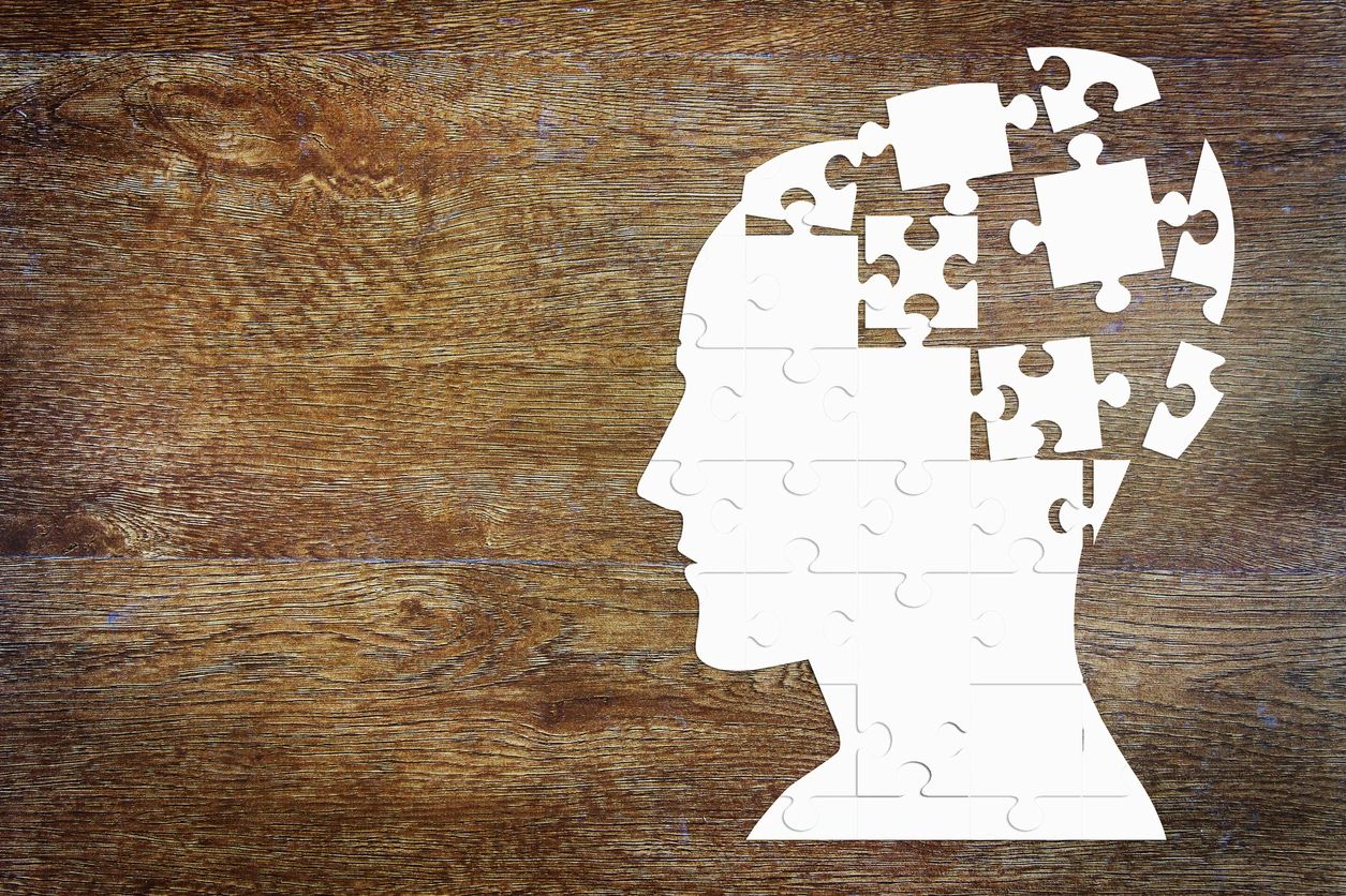

Panic attacks aren’t yet fully understood, but researchers have found a new neuronal pathway that may hold some answers.
For many, public speaking is the stuff of nightmares. It can give you sweaty palms, a rapid heartbeat, and even make you feel dizzy.
These are well-known symptoms of anxiety which are more physical than mental—what scientists call “somatic” symptoms. They serve to prepare the body for a coming threat. Sometimes they can explode in the form of panic attacks: periods in which a person experiences uncontrollable fear along with heart palpitations, sweating, trembling, shortness of breath, and similar symptoms.
Panic attacks are surprisingly common; every year, an estimated 11 percent of Americans have one. But when they become persistent, and don’t seem to have a cause (like that super-stressful work meeting or school project), it suggests that something is wrong with the body’s internal alarm bells. This is known as panic disorder, which affects 2–3 percent of Americans. The sudden, intense, and unpredictable burst of somatic symptoms sets panic disorder apart from other forms of anxiety.
What part of the brain controls panic attacks?
The brain structures that spark panic attacks are still unknown. One brain structure called the amygdala is famous (perhaps infamous?) for being the “fear center” of the brain. Particularly, it’s known for its role in fear conditioning: the process by which animals become afraid of something harmless, after it’s been associated with something harmful. For example, playing a neutral sound before giving a small shock to the foot of a mouse can make the mouse afraid of the sound. Hearing it, the mouse will freeze in place, anticipating a shock.
Since fear is central to a panic attack, it’s long been thought that the amygdala must mediate them. However, people who have had damage to their amygdalas (as a result of disease) can still experience panic attacks. This suggests that the amygdala isn’t the lone regulator. So, what other regions of the brain contribute? What more is there to panic attacks besides fear?
RELATED: Brain Circuits Map out Depression and Anxiety
The parabrachial nucleus: a little-known alarm center
Sukjae Kang’s research team at the Salk Institute for Biological Studies tried to answer this question, focusing on a small brain region known as the parabrachial nucleus (PBL). It’s located in the pons, a part of the brainstem. Some neurons within the PBL connect to the amygdala and are thought to be important for fear conditioning. The PBL also helps control some of the body’s subconscious functions, like breathing and heart rate regulation. As such, it could be a promising candidate for triggering panic attacks, where these functions run awry.
The researchers looked at a specific group of neurons in the PBL: those that produce a molecule called PACAP (aka PACAP neurons). This protein is known as a “master regulator” of stress responses. This makes PACAP neurons of the PBL prime suspects for mediating the onset of panic.
Kang’s team first asked what brain regions such neurons connect to. They used a virus which can travel across the synapses that connect neurons, carrying a fluorescent molecule that can be observed by microscope. They found that PACAP neurons of the PBL in mice connect not only to the amygdala, but also to another brain area called the dorsal raphe nucleus (DR). Neurons in the DR produce serotonin, a neurotransmitter involved in mood and sleep regulation, among other functions. Drugs targeting the serotonergic system in the brain have been used to treat anxiety and mood disorders. But what exactly are the consequences of this PBL-to-DR connection?
RELATED: Stress-Induced Sleep: A Built-In Snooze Button
How does a mouse panic?
Kang and colleagues wanted to understand the role of DR-projecting PBL neurons in controlling panic-like behaviors. First, they wanted to know if these neurons are activated when an animal is panicking. Brief exposure to elevated levels of CO2—though ultimately harmless—can readily cause a mouse to panic. They freeze, staying motionless, and their heart and breathing rates accelerate. The procedure has also been done on humans, to test new anxiety medications.
The researchers combined this procedure with calcium imaging—a form of microscopy that can visualize activated neurons in living animals. They found that the PACAP neurons of the PBL which project to the DR (PACAPPBL->DR neurons, for short) are more active during this CO2 challenge. The neurons were also activated by foot shocks as part of auditory fear conditioning, described earlier. Interestingly, however, these neurons weren’t activated when the conditioned sound was played on its own. In fact, the PACAPPBL->DR neurons were notably less active when the sound was played. This suggests that there are different pathways in the brain which generate panic versus conditional fear, and that PACAPPBL->DR neurons contribute only to the former.
To test this more directly, the researchers wanted to know if activating the neurons could itself cause the mice to panic. To do so, they used optogenetics: a cutting-edge tool which allows scientists to turn on neurons using light. A specific receptor that activates neurons in response to light, called channelrhodopsin, is delivered to the cells of interest—in this case, PACAPPBL->DR neurons. Then, a thin fiber-optic cable is used to transmit light from a laser or LED into the brain. Using this strategy, the researchers could selectively turn on the PACAPPBL->DR neurons in living mice.
They tested the role of PACAPPBL->DR neuron activation in fear conditioning, by playing a neutral sound paired with the neuron-activating light rather than a foot shock. Kang’s team found that the mice froze after the light activation, indicating panic. However, the mice didn’t freeze after hearing the sound on its own, again suggesting that PACAPPBL->DR neurons are not involved in fear conditioning.
Stopping panic in its tracks
Finally, the researchers wondered if inhibiting the PACAPPBL->DR neurons could reduce panic caused by the CO2 challenge from earlier. In this part of the study, they used the DREADD (Designer Receptors Exclusively Activated by Designer Drugs) approach. In this approach, receptors which are activated by a synthetic drug (in this case, hM4Di receptors, activated by the drug CNO) are delivered to cells of interest. Then, the deactivating drug can be injected into the animals, silencing the targeted neurons. When the researchers did this with PACAPPBL->DR neurons, the exposure to elevated CO2 didn’t panic the mice as much. They showed smaller spikes in heart and breathing rate, and less time spent immobilized.
RELATED: What happens to the brain in a good mood? Learn more about Brains and Play Behavior in Rats
Panic attacks, fear, and anxiety
Kang’s team conclude that conditioned fear, anxiety, and panic are different ways of responding to a stressor, and they rely on different pathways in the brain. Specifically, panic is encoded by PACAPPBL->DR neurons, while conditioned fear and anxiety probably rely more on connections between the PBL and amygdala.
In panic disorder, sufferers feel unconditioned fear; that is, their fear isn’t sparked by a stimulus, like a simple tone or bell. It originates out of nowhere, along with an intense wave of somatic symptoms. The activation of PACAPPBL->DR neurons in this study mirrors this pattern of symptoms. It causes an abrupt rise in heart and respiration rates, along with a spike in freezing behavior, without relying on a conditioned stimulus.
That isn’t to say that the amygdala has no role in panic disorder. In fact, Kang and colleagues suggest that agoraphobia (fear of open, public spaces), which often accompanies panic disorder, may point to an associative fear memory created by the amygdala: When an attack occurs outside of a “safe” place, it causes the individual to be afraid of such spaces.
RELATED: Anxiety Explained As an Evolutionary Response
However, the discovery of this new, “panicogenic” pathway in the brain opens up a much-needed new target for medications seeking to reduce panic attacks. Future research may help us figure out how other anxiety centers in the brain interact with the PBL, or what the DR does following stimulation by the PBL to generate panic-like behavior.
Until then, just take a deep breath and…well, try not to panic.
This study was published in the peer-reviewed journal Nature Neuroscience.
References
Cleveland Clinic. (2023). Panic Attacks & Panic Disorder. Retrieved January 23, 2024, from https://my.clevelandclinic.org/health/diseases/4451-panic-attack-panic-disorder
Feinstein, J. S., Buzza, C., Hurlemann, R., Follmer, R. L., Dahdaleh, N. S., Coryell, W. H., Welsh, M. J., Tranel, D., & Wemmie, J. A. (2013). Fear and panic in humans with bilateral amygdala damage. Nature Neuroscience, 16, 270–272. doi: 10.1038/nn.3323
Hammack, S. E., & May, V. (2015). Pituitary adenylate cyclase activating polypeptide in stress-related disorders: data convergence from animal and human studies. Biological psychiatry, 78(3), 167–177. doi: 10.1016/j.biopsych.2014.12.003
Kang, S. J., Kim, J. H., Kim, D. I., Roberts, B. Z., & Han, S. (2024). A pontomesencephalic PACAPergic pathway underlying panic-like behavioral and somatic symptoms in mice. Nature Neuroscience, 27, 90–101. doi: 10.1038/s41593-023-01504-3

About the Author
Rebecca DeGiosio is a recent PhD graduate in neurobiology from the University of Pittsburgh. She is soon to start a postdoctoral appointment at the Children’s Hospital of Philadelphia. Rebecca has a passion for translational biological research, particularly on psychiatric and neurodevelopmental disorders, and for making this research accessible to the public. Find her on LinkedIn: https://www.linkedin.com/in/rebecca-degiosio/